Category Archives: Symptoms

Batten Disease

What is Batten Disease?
Batten disease is named after the British pediatrician who first described it in 1903. Also known as Spielmeyer-Vogt-Sjogren-Batten disease, it is the most common form of a group of disorders called Neuronal Ceroid Lipofuscinoses (or NCL).

Although Batten disease is usually regarded as the Juvenile form of NCL, it has now become the term to encompass all forms of NCL.

The forms of NCL are classified by age of onset and have the same basic cause, progression and outcome but are all genetically different, meaning each is the result of a different gene. Over time, affected children suffer mental impairment, worsening seizures, and progressive loss of sight and motor skills. Eventually, children with Batten disease/NCL become blind, bedridden and unable to communicate, and, presently, it is always fatal.

Batten disease is not contagious or, at this time, preventable.

The History of Neuronal Ceroid Lipofuscinosis
The first probable instances of this condition were reported in 1826 in a Norwegian medical journal by Dr. Christian Stengel, who described three affected siblings in a small mining community in Norway. Although no pathological studies were performed on these children, the clinical descriptions are so succinct that the diagnosis of the Spielmeyer-Sjogren (juvenile) type is fully justified. More fundamental observations were reported by F.E. Batten in 1903, and by Vogt in 1905, who performed extensive clinicopathological studies on several families. Retrospectively, these papers disclose that the authors grouped together different types of the disease.

Furthermore Batten, at least for some time, insisted that the condition he described was distinctly different from Tay-Sachs Disease, the prototype of a neuronal lysosomal disorder now identified as GM2-Gangliosidosis type A. Around the same time, Spielmeyer reported detailed studies on three siblings, suffering from the Spielmeyer-Sjogren (juvenile) type, which led him to the very firm statement that this malady is not related to Tay-Sachs Disease. Subsequently, however, the pathomorphological studies of Schaffer made these authors change their minds to the extent that they reclassified their respective observations as variants of Tay-Sachs Disease, which caused confusion for about 50 years.

In 1913-14, M. Bielschowsky delineated the Late Infantile form of NCL. However, all forms were still thought to belong in the group of “familial amaurotic idiocies,” of which Tay-Sachs was the prototype.

In 1931, the Swedish psychiatrist and geneticist, Torben Sjogren, presented 115 cases with extensive clinical and genetic documentation and came to the conclusion that the disease which we now call the Spielmeyer-Sjogren (juvenile) type is genetically separate from Tay-Sachs.

Departing from the careful pathomorphological observations of Spielmeyer, Hurst, Sjovall and Ericsson, Zeman and Alpert made a determined effort to document the previously suggested pigmentary nature of the neuronal deposits in certain types of storage disorders. Simultaneously, Terry, Korey and Svennerholm demonstrated a specific ultrastructure and biochemistry for Tay-Sachs Disease, and these developments led to the distinct identification, and separation, of the NCLs from Tay-Sachs Disease by Zeman and Donahue. At that time, it was proposed that the Late Infantile (Jansky-Bielschowsky), the Juvenile (Spielmeyer-Vogt), and the adult forms (Kufs) were quite different from Tay-Sachs Disease with respect to chemical pathology and ultrastructure, and also different from other forms of sphingolipidoses. Subsequently, it was shown by Santavuori and Haltia that an Infantile form of NCL exists, which Zeman and Dyken had included with the Jansky-Bielschowsky type.

What are the forms of NCL/Batten Disease?
There are four main types of NCL, including two forms that begin earlier in childhood and a very rare form that strikes adults. The symptoms are similar but the forms become apparent at different ages and progress at different rates.

  • Infantile NCL (Santavuori-Haltia disease) begins between about 6 months and 2 years of age and progresses rapidly. Affected children fail to thrive and have abnormally small heads (microcephaly). Also typical are short, sharp muscle contractions called myoclonic jerks. Initial signs of this disorder include delayed psychomotor development with progressive deterioration, other motor disorders, or seizures. The Infantile form has the most rapid progression and children live into their mid-childhood years.
  • Late Infantile NCL (Jansky-Bielschowsky disease) begins between ages 2 and 4. The typical early signs are loss of muscle coordination (ataxia) and seizures along with progressive mental deterioration. This form progresses rapidly and ends in death between ages 8 and 12.
  • Juvenile NCL (Batten disease) begins between the ages of 5 and 8. The typical early signs are progressive vision loss, seizures, ataxia or clumsiness. This form progresses less rapidly and ends in death in the late teens or early 20s, although some may live into their 30s.
  • Adult NCL (Kufs disease or Parry disease) generally begins before the age of 40, causes milder symptoms that progress slowly, and does not cause blindness. Although age of death is variable among affected individuals, this form does shorten life expectancy.

There are six additional diseases included in the Batten disease/NCL group:

  • Finnish Late Infantile – identified in Finland
  • Variant Late Infantile – identified in Costa Rica, South America, Portugal and other nations
  • Turkish Late Infantile – identified in Turkey
  • Northern Epilepsy/ERMP – Epilepsy with Mental Retardation – identified in Finland
  • Variant Juvenile – identified in Germany and USA
  • Congenital/CTSD – identified in Europe

A more precise chart of the forms of Batten disease is below:

Chart: Forms of Batten Disease
Form Initials  Gene  Age of Onset 
Infantile INCL CLN1 6 mos. — 2 yrs.
Late Infantile LINCL CLN2 2 — 4 yrs.
Juvenile JNCL CLN3 5 — 7 yrs.
Adult ANCL CLN4 25 — 40 yrs.
Finnish Late Infantile fLINCL CLN5 2 — 4 yrs.
Variant Late Infantile vLINCL CLN6 3 — 5 yrs.
Turkish Late Infantile tLINCL CLN7 2 — 4 yrs.
Northern Epilepsy EPMR CLN8 5 — 10 yrs.
Variant Juvenile vJNCL CLN9 5 — 7 yrs.
Congenital CTSD CLN10 Birth — 2 yrs.

How many people have these disorders?
Batten disease/NCL is relatively rare, occurring in an estimated 2 to 4 of every 100,000 births in the United States, but no one really knows how many affected children there may be in North America or anywhere else in the world. The diseases have been identified worldwide. Although NCLs are classified as rare diseases, they often strike more than one person in families that carry the defective gene.

How are NCLs inherited?
Childhood NCLs are autosomal recessive disorders; that is, they occur only when a child inherits two copies of the defective gene, one from each parent. When both parents carry one defective gene, each of their children faces a one in four chance of developing NCL. At the same time, each child also faces a one in two chance of inheriting just one copy of the defective gene. Individuals who have only one defective gene are known as carriers, meaning they do not develop the disease, but they can pass the gene on to their own children.

Adult NCL may be inherited as an autosomal recessive (Kufs) or, less often, as an autosomal dominant (Parry) disorder. In autosomal dominant inheritance, all people who inherit a single copy of the disease gene develop the disease. As a result, there are no unaffected carriers of the gene.

What causes these diseases?
Symptoms of Batten disease/NCLs are linked to a buildup of substances called lipopigments in the body’s tissues. These lipopigments are made up of fats and proteins. Their name comes from the technical word lipo, which is short for “lipid” or fat, and from the term pigment, used because they take on a greenish-yellow color when viewed under an ultraviolet light microscope.

The lipopigments build up in cells of the brain and the eye, as well as in skin, muscle, and many other tissues. Inside the cells, these pigments form deposits with distinctive shapes that can be seen under an electron microscope. Some look like half-moons (or comas) and are called curvilinear bodies; others look like fingerprints and are called fingerprint inclusion bodies; and still others resemble gravel (or sand) and are called granual osmophilic deposits (GRODS).

Batten Disease - What causes these diseases?

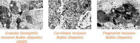
These deposits are what doctors look for when they examine a skin sample to diagnose Batten disease. The diseases cause the death of neurons (specific cells found in the brain, retina and central nervous system). The reason for neuron death is still not known.

How are these disorders diagnosed?
Because vision loss is often an early sign, Batten disease/NCL may first be suspected during an eye exam. An eye doctor can detect a loss of cells within the eye that occurs in the three childhood forms of Batten disease/NCL. However, because such cell loss occurs in other eye diseases, the disorder cannot be diagnosed by this sign alone.

Often an eye specialist/ophthalmologist or other physician who suspects Batten disease/NCL may refer the child to a neurologist, a doctor who specializes in diseases of the brain and nervous system. In order to diagnose Batten disease/NCL, the neurologist needs the patient’s medical history and information from various laboratory tests. Below are pictures of the retina showing the telltale signs of Batten disease.

Batten Disease and Ophthalmology

Batten Disease-How are these disorders diagnosed?

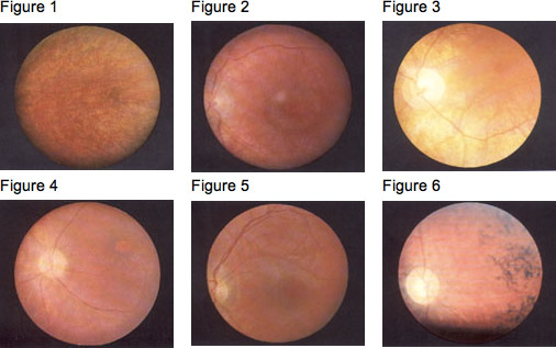
In the Fundus (the interior surface of the eye), the pigmentary changes in the macula are initially slight, and so it is easy to miss them, especially when no pupil dilation is applied and the fundus is not examined carefully. Fluorescent angiography demonstrates the pigmentary changes more clearly (Prammer, et al., 1978- ); sometimes fluorescence can be observed, leaking out of the retinal vessels. The density of the fine particuled pigmentations is slight around the macula and increases towards the periphery (Gottinger, et al., 1971- ). Dyken (1976) also mentions peripheral depigmentation. The pigment epithelium frequently has a granular “pepper and salt” appearance (see Fig. 1); sometimes there is a characteristic “bull’s eye” maculopathy (see Fig. 2, Fig. 3). The papilla becomes paler and the retinal arterioles seem more obviously constricted and extended (Fig. 4, Fig. 5). The peripheral retina varies in appearance, from normal to showing pigment-epithelial (pigmented cell layer just outside the retina) abnormalities (Spalton, et al., 1980- ). Later, peripheral pigment is often seen in the form of bone corpuscular pigment (see Fig. 6). Cataracts develop later in the course of the disease.

Diagnostic tests used for Batten disease/NCLs include:
Skin or Tissue Sampling: The doctor examines a small piece of tissue under an electron microscope. The powerful magnification of the microscope helps the doctor spot typical NCL deposits. These deposits are found in many different tissues, including skin, muscle, conjunctiva, rectal and others. Blood can also be used. See inclusion body pictures above.

Electroencephalogram or EEG: An EEG uses special patches placed on the scalp to record electrical currents inside the brain. This helps doctors see telltale patterns in the brain’s electrical activity that suggest a patient has seizures.

Electrical Studies of the Eyes: These tests, which include visual-evoked responses (VER) and electro-retinagrams (ERG), can detect various eye problems common in childhood Batten disease/NCLs.

Brain Scans: Imaging can help doctors look for changes in the brain’s appearance. The most commonly used imaging technique is computed tomography (CT), which uses x-rays and a computer to create a sophisticated picture of the brain’s tissues and structures. A CT scan may reveal brain areas that are decaying in NCL patients. A second imaging technique that is increasingly common is magnetic resonance imaging, or MRI. MRI uses a combination of magnetic fields and radio waves, instead of radiation, to create a picture of the brain.

Enzyme Assay: A recent development in the diagnosis of Batten disease/NCL is the use of enzyme assays that look for specific missing lysosomal enzymes for Infantile and Late Infantile only. This is a quick and easy diagnostic test.

Genetic/DNA Testing: Each “form” of Batten disease is the result of a different gene. Genes for eight of the ten forms have been identified. Testing for these is available for diagnosis as well as carrier and prenatal status.

Is there any treatment?
As yet, no specific treatment is known that can halt or reverse the symptoms of Batten disease/NCL. However, seizures can be reduced or controlled with anticonvulsant drugs, and other medical problems can be treated appropriately as they arise. At the same time, physical and occupational therapy may help patients retain function as long as possible.

BDSRA helps scientists by fostering awareness, promoting more research, providing samples and information, and by funding research that is directed towards understanding all forms of Batten disease and development of therapies.

Support and encouragement can help children and families cope with the profound disability and losses caused by NCLs. The Batten Disease Support and Research Association enables affected children, adults and families to share common concerns and experiences.

Meanwhile, scientists pursue medical research that will someday yield an effective treatment.

Trisomy 16

Chromosome 16 normally occurs in cells as a pair of chromosomes, one inherited from each parent. But when it comes to chromosomes, as the old saw says, anything that can go wrong, will. Pieces of the chromosome can mistakenly be duplicated, or may break off and get lost, or there can be too many copies of the entire chromosome. Below are some of the disorders of chromosome 16.

Trisomy 16
Instead of the normal pair, there are three copies of chromosome 16. Trisomy 16 is estimated to occur in more than 1% of pregnancies, making it the most common trisomy in humans. Unfortunately, this also makes trisomy 16 the most common chromosomal cause of miscarriages, as the condition is not compatible with life.

Trisomy 16 mosaicism
Sometimes there may be three copies of chromosome 16, but not in all cells of the body (some have the normal two copies). This is called mosaicism. Symptoms of trisomy 16 mosaicism include:

  • poor growth of the fetus during pregnancy
  • congenital heart defects, such as ventricular septal defect (16% of individuals) or atrial septal defect (10% of individuals)
  • unusual facial features
  • underdeveloped lungs or respiratory tract problems
  • musculoskeletal anomalies
  • urethral opening too low (hypospadias) (7.6% of boys).

There is also an increased risk of premature birth for infants with trisomy 16 mosaicism.

16 p minus (16p-)
In this disorder, part of the short (p) arm of chromosome 16 is missing. A disorder associated with 16p- is Rubinstein-Taybi syndrome.

16 p plus (16p+)
The duplication of some or all of the short (p) arm of chromosome 16 may cause:

  • poor growth of the fetus during pregnancy and of the infant after birth
  • small round skull
  • scant lashes and eyebrows
  • round flat face
  • prominent upper jaw with small lower jaw
  • round low-set ears with deformities
  • thumb anomalies
  • severe mental impairment.

16 q minus (16q-)
In this disorder, part of the long (q) arm of chromosome 16 is missing. Some individuals with 16q- may have severe growth and developmental disorders, and anomalies of the face, head, internal organs, and musculoskeletal system.

16 q plus (16q+)
Duplication of some or all of the long (q) arm of chromosome 16 may produce the following symptoms:

  • poor growth
  • mental impairment
  • asymetrical head
  • high forehead with short prominent or beaked nose and thin upper lip
  • Joint anomalies
  • genitourinary anomalies.

Charcot-Marie-Tooth disease

Charcot-Marie-Tooth disease, or CMT, is a group of inherited disorders that affect the peripheral nerves, which are the nerves outside the brain and spinal cord. There are more than 70 kinds of CMT. Each kind is caused by a different kind of mutation, and more causes are being discovered every year.

CMT is just one kind of neuropathy (also called peripheral neuropathy). These names simply mean that the peripheral nerves are damaged. There are many other causes of neuropathy besides CMT, including the most common cause—diabetes.

CMT affects all people, all races, and all ethnic groups, throughout the world, affecting about 2.8 million people!

Where Did the Name CMT Come From?
Charcot-Marie-Tooth is named after three physicians who were the first to describe it in 1886: Jean-Martin Charcot, Pierre Marie, and Howard Henry Tooth.

Inherited Disorders
Things in our environment do not cause CMT. CMT is not contagious. CMT is inherited. The most common forms of CMT are passed down from one generation to the next, meaning that it is dominantly inherited.

Some forms of CMT are recessively inherited—a person may be affected even though the parents do not have CMT. In this case, each of the parents harbors a mutation in one of their two copies of a CMT gene. If the child inherits the one mutated CMT gene from each of their parents (the chance of this happening is 1 out of 4), then the child will develop CMT.

Sometimes the mutation that causes CMT happens spontaneously during the process that produces the eggs or sperm. In these rare cases, a child will have CMT even though neither parent has CMT. If a child has such a spontaneous mutation, he/she may pass that mutation down to his/her offspring.

Symptoms
Some types of CMT cause damage to the covering (myelin sheaths) that surrounds nerve fibers. Other kinds of CMT directly damage the nerves fibers themselves. In both cases, the damaged nerve fibers result in neuropathy. The nerves in the legs and arms, which are the longest, are affected first. Nerve fibers that create movement (called motor fibers) and nerve fibers that transmit sensations (called sensory fibers) are both affected. Thus, CMT causes weakness and numbness, usually, starting in the feet.

In the most common kinds of CMT, symptoms usually begin before the age of 20 years. They may include:

  • Foot deformity (very high arched feet);
  • Foot drop (inability to hold foot horizontal);
  • “Slapping” gait (feet slap on the floor when walking because of foot drop);
  • Loss of muscle in the lower legs, leading to skinny calves;
  • Numbness in the feet;
  • Difficulty with balance;
  • Later, similar symptoms also may appear in the arms and hands.

CMT almost never affects brain function.

Diagnosis
A thorough neurological evaluation by an expert in neuropathy, including a complete family history, physical exam, and nerve conduction tests, with appropriate genetic testing is the way to establish the diagnosis of CMT.

A physical exam may show:

  • Difficulty lifting up the foot while walking;
  • Difficulty with dorsiflexion of the toes and ankles (upward movement, away from the ground) and other foot movements;
  • Reduced or absent deep tendon reflexes (like the knee-jerk reflex);
  • Loss of muscle control and atrophy (shrinking of the muscles) in the feet and lower legs (and later the hands).

Genetic testing can provide the exact cause for most people who have CMT.

Prognosis (Expectations)
CMT usually gets worse, slowly, with age; rapid progression is rare, and should motivate a prompt re-evaluation. The problems with weakness, numbness, difficulty with balance, and orthopedic problems can progress to the point of causing disability. Pain can be an issue, as a direct result of the neuropathy (neuropathic pain) and as consequence of orthopedic problems. Other potential complications include the following:

  • Progressive inability to walk from weakness, balance problems, and/or orthopedic problems;
  • Progressive inability to use hands effectively;
  • Injury to areas of the body that have decreased sensation.

Treatments
There are no known treatments that will stop or slow down the progression of CMT, but the CMTA is funding research to find these treatments.

Physical therapy, occupational therapy, and physical activity may help maintain muscle strength and improve independent functioning.

Orthopedic equipment (such as braces, inserts, or orthopedic shoes) may make it easier to walk.

Orthopedic surgery on the feet can often maintain or even restore function to enable walking.

Cat Eye Syndrome / Schmid Fraccaro Syndrome

Cat Eye Syndrome is the more common name for a condition involving a partial trisomy or tetrasomy of part of chromosome 22. A small extra chromosome (humans normally have only 2) made up of the top half of chromosome 22 – the “p” arm, as well as the portion of the long arm of chromosome 22 down to the breakpoint q11.2, is found to be present either three times (trisomy) or four times (tetrasomy ).

Cat Eye Syndrome is also known in the literature as Schmid-Fraccaro Syndrome, Partial Tetrasomy 22, or Inv Dup(22)(q11) (Inverted Duplication).     It is often referred to as Cat Eye Syndrome as some of the people affected may have coloboma of the iris – which make their eyes appear to look like cat’s eyes. This feature however, is only reported in about half of the known cases. The earliest reports stem back over 100 years ago, but the first association

Clinically, this is one of the more variable syndromes. People who have Cat Eye Syndrome can be anywhere from normal to suffering from severe malformations. This has proven true of the people who have joined our group, who have this condition.

Some of the features seen in people with CES include coloboma of the iris, anal atresia, ear tags and/or ear pits, heart defects, and kidney malformations, but because of the variability, there have been reports of malformations affecting almost every organ.

Cognitively, people with CES can be considered either normal intelligence, or have varying degrees of mental delay, although it is rare to see severe mental impairment in this condition.

The condition usually arises spontaneously, but our group does have some members who have CES and have passed it on to their children.

Trisomy 18/Edwards syndrome

Trisomy 18, also known as Edwards syndrome, is a condition which is caused by a error in cell division, known as meiotic disjunction.  Trisomy 18 occurs in about 1 out of every 2500 pregnancies in the United States, about 1 in 6000 live births.  The numbers of total births increase significantly when stillbirths are factored in that occur in the 2nd and 3rd trimesters of pregnancy.

Unlike Down syndrome, which also is caused by a chromosomal defect, the developmental issues caused by Trisomy 18 are associated with medical complications that are more potentially life-threatening in the early months and years of life. 50% of babies who are carried to term will be stillborn, with baby boys having higher stillbirth rate than baby girls.

At birth, intensive care admissions in Neonatal units are most common for infants with Trisomy 18. Again, baby boys will experience higher mortality rates in this neonatal period than baby girls, although those with higher birth weights do better across all categories.

Some children will be able to be discharged from the hospital with home nursing support for their families. And although less than 10 percent survive to their first birthdays, some children with Trisomy 18 can enjoy many years of life with their families, reaching milestones and being involved with their community.  A small number of adults (usually girls) with Trisomy 18 have and are living into their twenties and thirties, although with significant developmental delays that do not allow them to live independantly without assisted caregiving.

What causes Trisomy 18?
At conception, 23 chromosomes from the father and 23 chromosomes from the mother combine to create a baby with a set of 46 chromosomes in each cell. A trisomy occurs when a baby has three #18 chromosomes instead of the normal two. This is something that happens at conception. And although many parents worry about this, it is important to know that parents have done nothing before or during pregnancy to cause this disorder in their child.

Are there different types of Trisomy conditions?
The most common trisomy is Trisomy 21, also known as Down syndrome, where a baby has three of the twenty-first chromosome. Trisomy 18 is the second most common trisomy and occurs when a baby has three of the eighteenth chromosome. This results in 47 chromosomes instead of the normal 46 in the affected cells. It is this extra genetic material that causes the problems associated with Trisomy 18. The third most common is Trisomy 13, also known as Patau syndrome.

While there are different types of Trisomy 18, this does not mean one is better for a child than another.  With each type, there is a range of possibilities. Some children are medically fragile while others thrive; some children walk while others are confined to wheelchairs. It is hard to say how the extra chromosome will impact an individual child from the genetic diagnosis alone.

Types of Trisomy 18:

  • Full Trisomy 18: The most common type of Trisomy 18 (occurring in about 95% of all cases) is full Trisomy. With full Trisomy, the extra chromosome occurs in every cell in the baby’s body. This type of trisomy is not hereditary. It is not due to anything the parents did or did not do—either before or during pregnancy.
  • Partial Trisomy 18: Partial trisomies are very rare.  They occur when only part of an extra chromosome is present. Some partial Trisomy 18 syndromes may be caused by hereditary factors. Very rarely, a piece of chromosome 18 becomes attached to another chromosome before or after conception. Affected people have two copies of chromosome 18, plus a “partial” piece of extra material from chromosome 18.
  • Mosaic Trisomy 18: Mosaic trisomy is also very rare. It occurs when the extra chromosome is present in some (but not all) of the cells of the body.  Like full Trisomy 18, mosaic Trisomy is not inherited and is a random occurrence that takes place during cell division.

What are the characteristics of Trisomy 18?
The genetic material from the extra eighteenth chromosome can cause a variety of problems with varying severity. Just as children with Down syndrome can range from mildly to severely affected, the same is true for children with Trisomy 18. This means that there is no hard and fast rule about what Trisomy 18 will mean for your child. However, statistics show that there is a high mortality rate for children with Trisomy 18 before or shortly after birth.

Typical characteristics of Trisomy 18 include:

  • Heart defects:
    • VSD (Ventricular Septal Defect): a hole between the lower chambers
    • ASD (Atrial Septal Defect): a hole between the upper chambers
    • Coarctation of the aorta: a narrowing of the exit vessel from the heart
  • Kidney problems
  • Part of the intestinal tract is outside the stomach (omphalocele)
  • The esophagus doesn’t connect to the stomach (esophageal artesia)
  • Excess amniotic fluid (polyhydramnios)
  • Clenched hands
  • Pocket of fluid on the brain (choroid plexus cysts)
  • Rocker bottom feet
  • Delayed growth
  • Small jaw (mycrognathia)
  • Small head (microcephaly)
  • Low-set ears
  • Strawberry-shaped head
  • Severe developmental delays
  • Umbilical or inguinal hernia